
Magnetic Resonance Imaging
Magnetic Resonance Imaging (MRI) is a powerful technique, suitable foremostly for imaging of soft tissues. It allows acquisition of anatomical as well as functional information in diverse research areas, including neurology, cardiology and oncology.
MRI is widely used as a non-invasive diagnostic imaging tool on a daily basis in hospitals and research centers. It has gained popularity in the preclinical imaging community for the wide range of applications where MRI can be employed
The Preclinical Research Center (PRC) is equipped with a variety of different imaging techniques, among these the magnetic resonance imaging scanner, a cryogen-free 3T Bruker Biospec has been installed in late 2019 in the PRC and it is primarily managed by DVM Dr. Ielacqua, Giovanna Diletta.
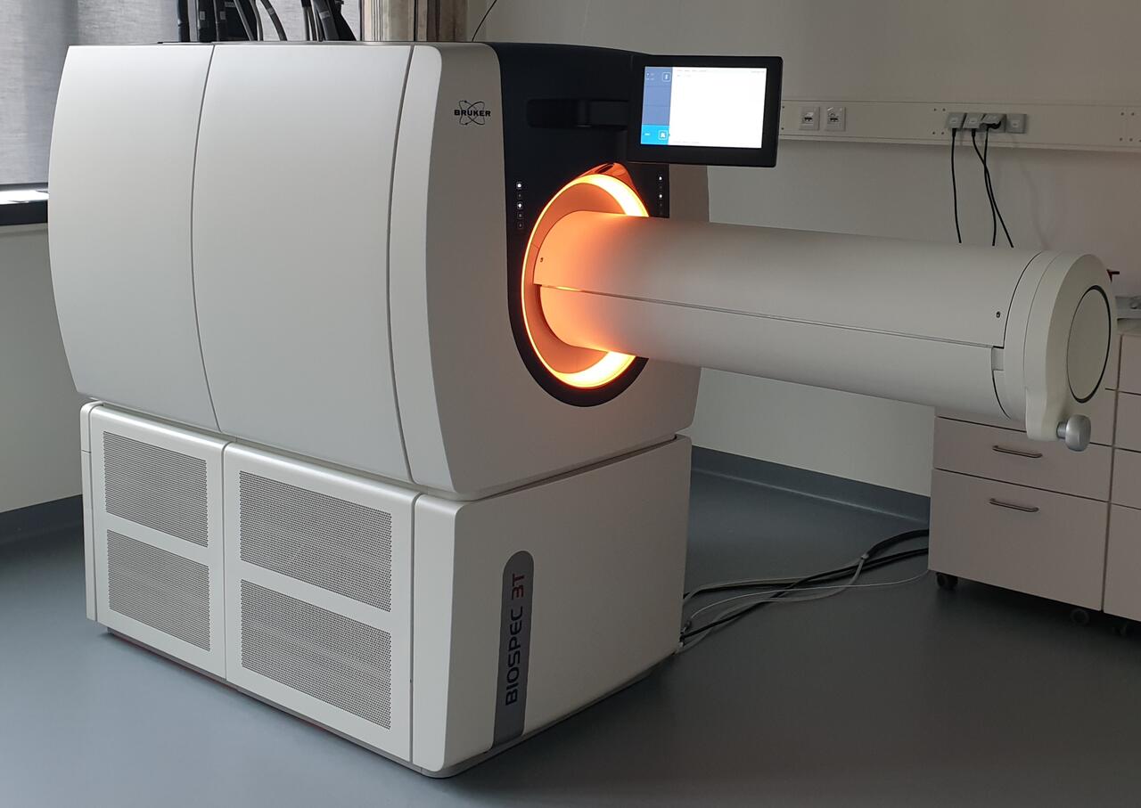
Our compact cryogen-free scanner operates at a translational field of 3 Tesla, the BioSpec 3T is one of the newest scanner available on the market and offers a wide range of different applications from preclinical imaging (MRI) to magnetic resonance spectroscopy (MRS). Its 180 mm bore allows to image not only mice and rats, but also other small rodents. The cryogen-free technology ensures a 4 hours magnet field stability during power and water outage and the self-shielded system does not require a Faraday cage.
The Bruker 3T BioSpec scanner available at our platform is equipped with diverse volume as well as surface coils for optimal image acquisition in numerous applications. Moreover, the motorized animal positioning system allows an easy and convenient performance of experiments.
Applications
The main preclinical applications comprise not only structural but also functional measurements in different fields such as neuroimaging, oncology, cardiology, cardiovascular and metabolic research allowing us to perform measurements in different organs as well as in joints, bones and whole-body imaging.
Features
The scanner operates with the latest Bruker software application packages Topspin and Paravision 360 which are already fully compatible with the MRI CryoProbe (which increases the signal-to-noise ratio to a factor of 2.5) and also other imaging modalities such as PET; The scanner is also equipped with a motorized animal handling system, including touchscreen operation for a better workflow to give a time-efficient and accurate animal positioning.
Our MRI facility includes also a state-of-the-art anesthesia monitoring system integrated into the MR scanner: cardiovascular parameters such as heart rate, pulse distension, Oxygen saturation, breathing rate and body temperature can be efficiently monitored in real time while animals are being scanned, allowing us to run measurements while ensuring that animal physiology and animal welfare are guaranteed.
In regard to data analysis the MR facility of the PRC offers a wide array of tools, ranging from more traditional imaging software such as FIJI and MatLab built-in scripts to more application-specific packages like FSL and SPM for functional neuroimaging. New software are constantly being tested and validated for potential future use and MR trainers/manager are regularly attending MR conferences and training courses to ensure that any new development in the MR field is taking into account and implemented when possible into the PRC MR facility.