
Buch campus to get ultralow temperature microscopes
If you want to make microscopically small structures like proteins visible at the nanometer level, you need electron beams to enlarge them. It also has to be very cold. Using cryo-EM, proteins are studied at temperatures of less than −150°C, allowing their three-dimensional structures to be imaged with a precision of 0.2 nanometers. In this way they retain their original form almost completely. This is important for analyzing their functionality.
In structural biology, we want to work very closely with Charité and other university partners in Berlin
In mid-December, two cryo-EMs belonging to Charité – Universitätsmedizin Berlin were delivered to the Buch campus in large crates. Until their assembly begins in February, they are being stored in their own special building at the Max Delbrück Center for Molecular Medicine in the Helmholtz Association (MDC). “We’re building a small house for a very large microscope,” commented Ralf Streckwall, head of construction for the Technical Facility Management Department, at festivities in November. The research building will be completed in the spring of 2020. The building alone will cost about €2.75 million. The largest microscope is nearly four meters high and is worth €5 million. Charité received a co-financing grant from the German Research Foundation (DFG) for 50 percent of the equipment costs.
Proteins in their cellular environment
Researchers in Berlin will probably be able to use the state-of-the-art technology for their projects by the middle of the year. “In structural biology, we want to work very closely with Charité and other university partners in Berlin,” says Dr. Jutta Steinkötter, head of Scientific Infrastructures at the MDC. “But there’s still a lot to do before that can happen,” adds Dr. Claudia Flügel, an expert on research infrastructure at Charité. “We’re pleased that Dr. Christoph Diebolder will be taking over as director of the Cryogenic Electron Microscopy Core Facility. Dr. Diebolder will assume that role on February 1, 2020.”
Many scientists at the MDC can’t wait to get started. Structural biologist Prof. Oliver Daumke is one of them. He studies proteins that perform important functions inside cells by remodeling cellular membranes during energy consumption. Admittedly, Daumke could illustrate their three-dimensional structures using crystallography, but that would only produce the image of one isolated protein. “What’s special about cryogenic electron microscopy,” Daumke explains, “is that it lets us look at proteins in their cellular environment.” Daumke also emphasizes that the new technology soon to be available at the Buch campus will be attractive for early career scientists in the process of developing their own research projects. The MDC is currently putting together a junior research group in this field.
Every vibration is a disturbance
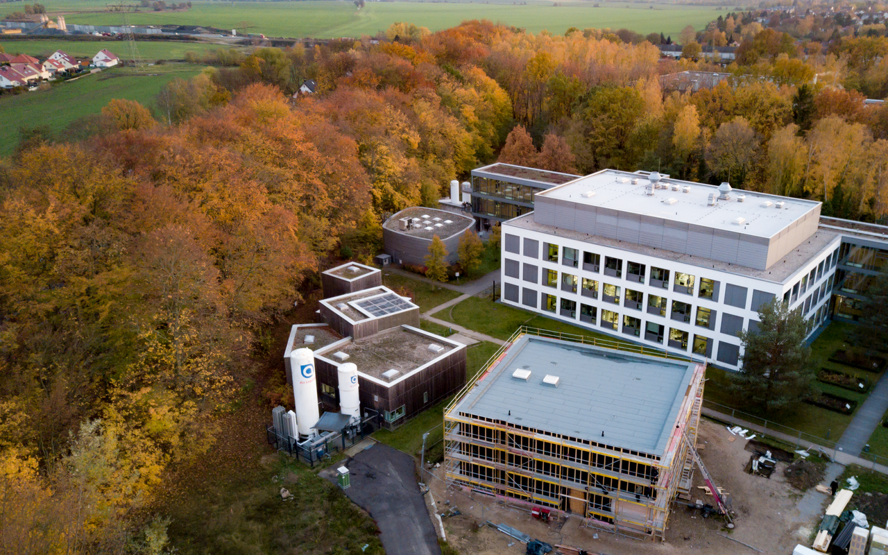
A new building for two Cryo-EM will be finished soon at the Buch campus.
It wasn’t easy to find a suitable location for the cryo-EMs in Berlin. According to Karsten Hönig, a project leader at the MDC, electromagnetic fields and underground vibrations are a problem. “Even the campus bus creates vibrations when it drives by that would impair the microscopes’ function. A concrete foundation 1.25 meters thick has therefore been placed underneath the building. Like a big, heavy ship on the water, it cancels out the vibrations of ground waves.”
The new building for the microscopes is only one of several construction projects currently underway at the Buch campus. Very close by, the MDC’s 2,700-square-meter Optical Imaging Center (OIC) is being built. The OIC is slated to be finished by 2020 and will enable a synergy between research, applications, and innovations in microscopy, (bio)physics, and the life sciences. It will be available to internal and external researchers, visiting scientists at the MDC, and partners from industry.


