
Globalizing medical imaging
An open-source project launched by an MDC postdoc aims to make MRI affordable and accessible world-wide.
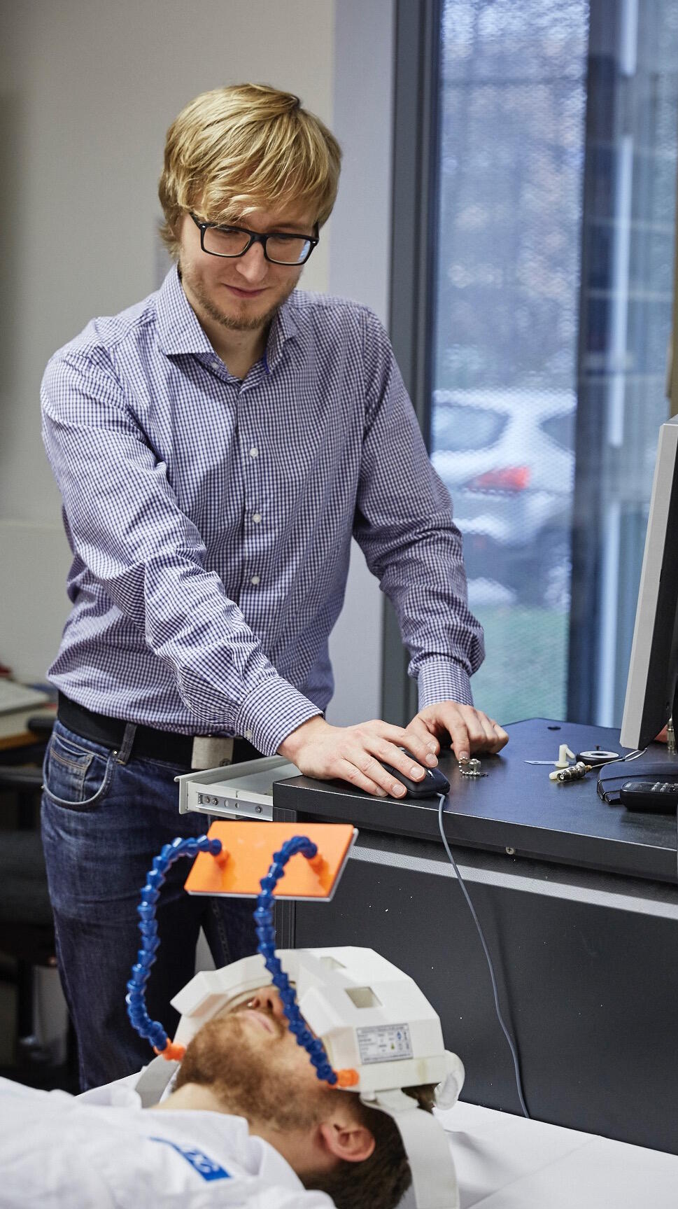
Lukas Winter, a postdoc in Thoralf Niendorf's lab, has a vision. Lukas is an expert in Magnetic Resonance Imaging (MRI), a technology that permits doctors and scientists to remotely view patients' internal organs and tissues without invasive surgical procedures. Now Lukas is activating the MRI community in an Open Source project to build instruments that will be affordable and accessible to people all over the world.
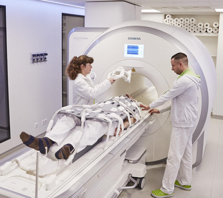
Lukas' work in the Niendorf lab has focused on “ultrahigh”-field MRI machines that use huge, superconductive magnets immersed in extremely cold liquid helium. The machines found in clinics throughout the industrial world are somewhat smaller, but are still extremely expensive to buy and maintain. This has made them inaccessible or very rare elsewhere, to the detriment of patients across the world.
While working on his PhD in the Niendorf lab, Lukas hit on a plan to make MR machines and their maintenance much more affordable: by enlisting the efforts of MR experts world-wide in the design of an "open-source" instrument. After months of careful planning, he presented the concept at a global MR conference in Singapore in May. The response was very enthusiastic, and scientists are joining working groups to tackle aspects of the design and other parts of the project. In this interview Lukas describes the idea and the steps that will be necessary to deliver working MR machines to patients and clinicians around the world.
MDC: You are a postdoc at the MDC. What is your professional research about?
Dr. Lukas Winter: I am working on the development of ultrahigh field MR hardware and methodology at a magnetic field strength of 7 Tesla. Such high-end instruments have many research applications and will reach many clinics in the next few years. But they are very expensive machines. Even the current clinical MR workhorses, which operate at field strengths of 1.5 T and 3.0 T, are unaffordable for many people around the world. Scanner acquisition costs are between 1 and 2.5 Mio Euros depending on the field strengths plus maintenance and operational cost of nearly half a million Euros annually.
That's really expensive. How would you reduce these hardware costs?
Technically, the most expensive part of a modern MR machine is the huge electromagnet, which also has high maintenance costs. The magnets are made of superconductors that have to be cooled with expensive liquid helium. They are turned on 24 hours a day, have to be constantly monitored, and require a dedicated room.
Our current plan involves using a number of small magnets which will be permanently mounted in a specific configuration – we're talking about magnets only a little stronger than the type you attach to your refrigerator! No external power and no helium needed! We will also use these arrangements to generate a magnetic field gradient that can be used for spatial encoding. This allows us to waive some of the classical homogeneity requirements of a magnet and to waive expensive gradient power amplifiers and gradient coils.
Using such magnets at low field of 0.2 Tesla allows to reduce the material costs for a complete MRI scanner including electronics to around 10.000 €.
So these ultrahigh fields are not always necessary?
Exactly. In many cases you don’t need a sledgehammer to crack a nut. MRI based on smaller magnetic fields can be extremely useful in diagnosing a range of health problems. While very high fields have advantages such as increased resolution and sensitivity, the physics of magnetic resonance reveals that smaller magnets have advantages of their own.
For example, lower fields are much safer. The attraction force of a weaker magnet is smaller, which is beneficial for patients that have metallic or magnetic implants, and there's a lower risk that magnetic objects such as pens, chairs or car keys might hurt the patient during an MR exam. The instruments will be smaller and quieter, which is a crucial consideration when dealing with newborns or children. The size of the instrument could permit taking one to a clinic somewhere in the field – bringing it to patients, rather than bringing them to the clinic. That is impossible with a large machine. There are probably lots of other possibilities that haven't really been explored.
You mentioned hardware costs are just one part of the equation. How would you reduce the "other" costs of MR imaging?
Jim Pipe, the president of the International Society for Magnetic Resonance in Medicine (ISMRM), recently stated: “…there are really four costs to total ownership [of an MR device]. There’s the scanner, the maintenance, the staffing, and then everything else – including power.”

An open source approach can address all these points. The open source concept comes from software design, where whole communities of people work on the source code of programs, to make something that anyone can use. The same approach might be applied to medical engineering. Sharing and collaboration are scientific values. Open source software and hardware go a bit farther than the rest of science by making not only the publication and data accessible, but also the code or the design of a machine, so that it can be accessed and modified by users.
The costs for scanner hardware can also be reduced by creating new component designs and outsourcing research and development from companies to the community. The key to reducing maintenance costs is very effective, transparent documentation. That's also a way to train staff and newcomers in the field like students, providing we reduce the complexity of the documentation and think about education from the beginning. We think the solution to "everything else" will come through the vast knowledge and creativity of the research and maker community. We'd be asking MR experts around the globe for a bit of their time and expertise and to make their own designs freely available toward this larger social goal – which is at the heart of all science, really.
But documentation in Open Source projects is often a mess – people hate to spend their spare time on boring but important stuff.
Exactly, just like in science. Scientists spend a lot of time on documentation like writing papers and grant applications don't they? And that’s the trick. A scientist's currency is publications or citations. A journal editor or peer reviewer can motivate the authors to publish their research open source (i.e. providing all necessary code or hardware schematics to reproduce the results) and document it in an understandable way. In exchange the scientist gets a publication. Even more so, fellow scientists are able to reproduce the results more easily and often cheaper which accelerates science, just like the open access movement does. Open source and science is a perfect marriage.
If the community designs it, who will decide which route to take, will there be a project leader or a chief designer?
I am coordinating the developments of the Open Source MR scanner in Buch. But in general there will be multiple project leaders or designers depending on the target application and complexity. Do you want to build an educational device, a research instrument or are you interested in building a device for a particular clinical application? Each scenario will have different requirements and specifications.
It is difficult for a single person or lab to build a complete system from scratch. If you split an MR scanner in different modules it is a different story. You have experts for RF power amplifiers, for RF coils, for gradient coils, for magnets, for spectrometers etc. You just need to bring this expertise together, make the modules transparent and define the interfaces. By access to information the true creativity can be unleashed. The best examples are 3D printers. Without them being open source we wouldn’t see such a variety of applications from printing household items to printing food, houses or even fully functional robots.
Once the initial stage of technological developments and proof of principle studies is complete, clinical questions will arise. At this point the commercial interest will increase, which is good and necessary for a dissemination of the technology. When transitioning from a research device or prototype to a medical device, quality, reliability and safety for the target application need to be ensured, which often requires resources from industry. With the development outsourced, the investment to enter a market can be lower. So basically if the demand for affordable and portable MR machines is there, which I firmly believe, we will see more competition and lower prices for the devices as well as for service contracts.
Obviously with a community project, communication will be crucial. How does this idea of building an open-source MR instrument become a practical reality?
Friends and I have dedicated a lot of our spare time building a collaboration hub, OpenSourceImaging.org. The website is part of the “Open Source Imaging Initiative (OSI²)”, which was co-authored by scientists from the Charité, Stanford, MIT, MGH, PTB, LUMC, University of Mainz and the TU-Berlin.
We presented the initiative a few weeks ago to the community at the meeting of the International Society for Magnetic Resonance in Medicine (ISMRM) in Singapore, the biggest conference related to MR technology. The feedback was amazing. We hope to keep discussion alive and make an impact on the community.

As far as I know this approach hasn't yet been used to build medical instruments on that scale. If we can do this, we might even be making a model that others can follow, to produce a range of other medical technologies more cheaply and make them more widely accessible. This might also work for a range of other complex medical and scientific problems.
What's your timeline? Where is the project at right now?
We are currently building a prototype magnet and finalizing the electronics in hopes of getting a first signal by the end of the year. Next year we aim to obtain our first image – cross your fingers!

We're now in a proof-of-principle phase in which we have already attracted the interest of labs and researchers across the world. We're getting advice on legal and regulatory issues and application procedures as well as communication aspects of the project. Thoralf Niendorf has been extremely supportive – even though this isn't a focus of the lab, he absolutely sees the sense in what we're trying to do. He understands that an important by-product of this type of work will be to produce important new research insights into the specific technology and its applications. Felix Arndt and Claudia Thurow from the legal department and Dr. Jeanette Libera-Körner from the technology transfer department at the MDC have been extremely supportive of the project, for example by helping to shape a workflow on the open source hardware license that will be used as well as providing legal advice on liability issues.
We have been identified as an “impressive change agent” by the Shuttleworth Foundation, which supports social innovators for an open knowledge society, and received a Flash Grant for the project. I also recently joined the editorial board of HardwareX, a new open access journal that promotes free and open-source designs for scientific infrastructure, which is tied to the Open Science Framework. So in general things are moving rapidly forward. It's exciting.

Challenges will appear, naturally, but my experience with research projects suggests there is always a solution. No one doubts that building MRI instruments is feasible, and no one doubts that the open source software movement has taken off to become a multi-billion dollar market today. The only limiting factor in determining how fast we can reach our goal will be the level of resources that we can attract in the process.
I think it's the right time for a project like this, and the people we have talked to are very enthusiastic about it. As I said, it's highly motivating. Part of that is a real need: statistics from the World Health Organization show that in terms of machines per capita, there is a difference by a factor of 100 or more in access to MR across the world – in countries that have it. Some parts of the world have no instruments or trained operators at all. Many sites are frustrated because existing machines can't be repaired. Even in developed countries, there are great differences in access; waiting times for an appointment in France are often much longer than a month, which can make a huge difference in the course of a disease and a treatment regime. We think that these are all problems which can be solved.
###
For more information about the project, and ways to participate, visit: www.opensourceimaging.org or write an email to Lukas.winter@mdc-berlin.de.
Featured image: Katharina Bohm/MDC.


