
The image-maker

Thoralf Niendorf
What does an opera singer have in common with medical imaging? According to Professor Thoralf Niendorf, plenty! The physicist researches potential uses of ultrahigh field magnetic resonance imaging (UHF-MRI). The magnetic field that this technology produces is far stronger than the MRI scanner you might know from hospitals. “At first, there were major doubts whether ultrahigh field devices were even suitable for heart examinations,” he recalls. After all, the beating heart is constantly in motion.
In order to record movement, an MRI would normally measure the electrical impulse of the heart muscle via an electrocardiogram (ECG). But at a magnetic field strength of 7.0, the interference is so strong that the MRI system is unable to recognize ECG signals with any clarity. “Thus, researchers all around the world were trying and failing to clearly image the heart with 7-Tesla MRI techniques,” explains Niendorf. That’s when opera came into play.
Niendorf was working with a team from the field of phoniatrics – the study and treatment of voice, speech and language disorders – when the idea occurred to him. They needed MRI images of the vocal chords. “We put professional opera singers in the MRI scanner and asked them to perform arias while we measured the organs involved in creating voice,” Niendorf explains. The “aha” moment came in discussion with a phoniatrics expert: acoustic signals don’t influence the magnetic field. The sounds that the heart makes are therefore perfect for visualizing the heart while in the MRI scanner. So, together with his team, he developed new high-frequency antennas – also known as coils – and created the world’s first clear 7-Tesla images of the human heart.
Visualizing cellular biological processes
The best ideas arise from the exchange with other disciplines.
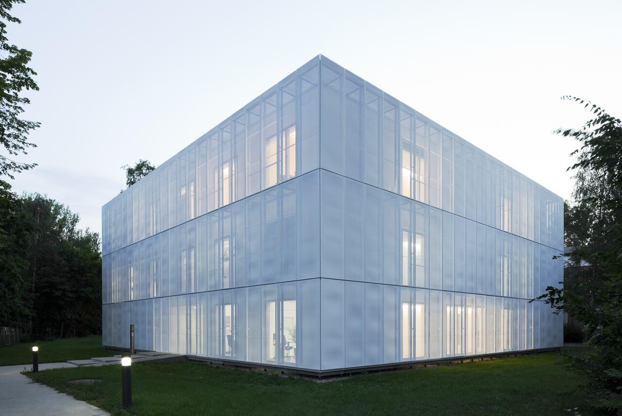
“The best ideas arise from the exchange with other disciplines,” says Niendorf, who heads the Experimental Ultrahigh Field MR Lab. He prefers to sort his thoughts when jogging or with water sports. Office talks, he finds, are rarely inspiring. So he suggests we hold the interview outside, sitting on the lawn in front of the white cube which is home to the Berlin Ultrahigh Field Facility (B.U.F.F.). The building houses clinical MRI devices with magnetic field strengths of 3 and 7 Tesla, as well as one with 9.4 Tesla that is not for use on humans. B.U.F.F. is a world-class facility that enables world-class research. Niendorf and his team are not only pushing the boundaries of anatomical imaging resolution, they’re developing new techniques to visualize physiological and cellular biological processes. As such, they open up completely new avenues for basic research but also for the early diagnosis and therapy of disease.
But one thing after the other: magnetic resonance imaging, which is based on the principles of nuclear resonance, has been around since the early '70s. Alternating electromagnetic fields excite atomic nuclei in a powerful magnetic field, and images of the spatial distribution of the nuclei can be calculated from data on the reception and emission of these signals. It is an imaging method that produces precise anatomical slice images without having to employ ionized radiation.
The first clinical MRI devices operated at a magnetic field strength of about 0.5 Tesla, while the standard hospital scanner now has between 1.5 and 3.0 Tesla. Among other things, the two 3.0 Tesla devices at B.U.F.F. are used to create whole-body scans of approximately 6,000 subjects taking part in the German National Cohort (or NAKO in German) health study. These highly detailed scans, together with anonymized data on things like blood tests, chronic illnesses, nutritional habits and lifestyle, are made available to the international research community.
The sky’s the limit
Aside from health research, Niendorf is particularly interested in new methodologies for ultrahigh field MRI. “Image resolution increases proportionately with an MRI’s field strength, Niendorf explains. The researcher removes all magnetic items from his pockets before opening the door to the centerpiece of the building: the 7.0 Tesla scanner. When he began his research on ultrahigh field MR imaging, there were only about 10 such devices in the entire world. The advantages that the increased resolution offered neurology quickly became evident, however. Today, about 100 clinics rely on the 20-ton 7.0 Tesla MRI as a brain scanner.
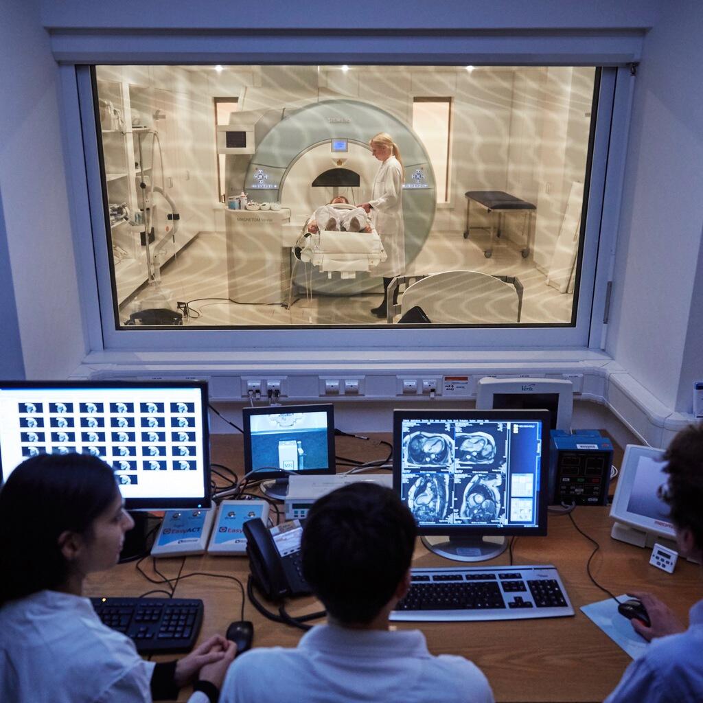
Scientists performing an MRI experiment.
And MRI devices with even greater field strength have long since been developed. “Technically, there is no limit to what can be done,” says Niendorf. It’s easily imaginable to have MRI images of individual cells. In experimental studies with the 9.4 Tesla device, structures as small as 20 micrometers have been clearly imaged.
Currently, the lab’s researchers are using the device to optimize kidney diagnostics. The project is part of the Collaborative Research Center “Renoprotection” at Charité – Universitätsmedizin Berlin. The extreme resolution of the images is uncovering fascinating details from within peanut-sized mouse kidneys. For example, they reveal oxygen deficiency very early on, indicating the risk of kidney injury. This is, of course, highly relevant as patients in intensive care units (ICU) often suffer acute kidney failure: one in three ICU patients develop the condition. “With high-resolution MRI images, we will be able to recognize deterioration of the kidney functions much earlier, and so save lives,” says Niendorf. The next step is to conduct human experimental studies with the 7.0 Tesla scanner.
Fighting tiny tumors with helmet-like coils
Niendorf takes out an MRI coil. It roughly resembles a bucket-shaped knight’s helmet, only this one is made not of iron but creme-tinted white plastic. “With this and a 7.0 Tesla scanner, I can examine an eyeball in incredible detail,” says Niendorf. His team developed it together with physicians at the University of Rostock. The high-resolution images of the eye produced with this technology revealed tiny tumors that loosen the retina and impair the patient’s vision. “These tumors were not visible to a clinical 3.0 Tesla scanner,” says Niendorf.

Thoralf Niendorf
Niendorf finds it even more interesting that the high field strengths can open up completely new methods of image generation. Commonly available MRI devices predominantly excite hydrogen nuclei in the field, creating an image from the measured tissue density. The higher field strengths allow direct representation of elements like sodium, potassium, phosphorus and fluorine. Each plays an important role in metabolism and so generate new ways of visualizing physiological processes.
Fluorine, for example, builds up in inflamed tissue. Therefore, it could be used as a biomarker for the early diagnosis of inflammation. The sodium-potassium pump in a cell’s membrane maintains a balanced concentration of both elements, which is crucial to the healthy functioning of neurons and most other cells. In cooperation with the German Cancer Research Center (DKFZ) in Heidelberg, Niendorf’s team was able to develop a technique to identify sodium ions in heart scans. Researchers can thus distinguish between healthy and damaged tissue in a beating heart – one of only many conceivable ways that this technology could be used to improve diagnostics in the future.
Homemade surfboards
Berlin is a great location for the sciences. No matter the field, there are partners out there.
How did he find his calling? Niendorf grew up in the East German town of Jüterbog, about 50 kilometers south of Berlin. From the beginning, his father encouraged him to question conventions and ideology. His parents excused him from school once a month so that he could visit classical concerts and theatre performances. When windsurfing became popular in the West, father and son made their own surfboards – and tested them out on the lakes of Brandenburg.
The sport continues to fascinate him to this day, and he can regularly be found at the Baltic Sea with his daughters, out on the water. “It helps me clear my head. I can structure my thoughts and ideas,” says Niendorf. His successful application for an ERC Advanced Grant was partially conceived and written on a sandy camping table on the Baltic coast. He also hasn’t stopped questioning accepted truths.
Today he’s happy to have followed the call to Berlin – after having been at the Max Planck Institute in Leipzig, at GE Healthcare in the US and at the RWTH Aachen University as a professor. “Berlin is a great location for the sciences. No matter the field, there are partners out there,” he says. He enjoys working in an environment where he’s comfortable with the nuances of communication and where he can further develop his network. Among his many ventures, the industrious scientist has founded the “MDC-Weizmann Helmholtz International Research School for Imaging and Data Science from the NAno to the MESo” and has played an instrumental role in establishing Helmholtz Imaging.
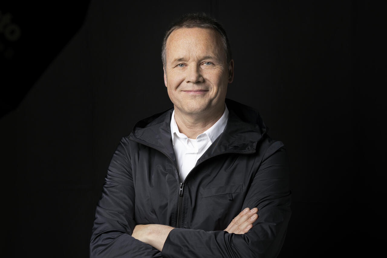
Thoralf Niendorf
Yet bringing the imaging sciences together with the life sciences still remains a challenge. “You first have to establish a mutual language and learn to understand the other side.” Then, however, good things happen: you formulate completely new questions – and find inspiration.
Text: Dietrich von Richthofen


